Fungal keratitis tends to occur after corneal injury involving plant material or in an agricultural setting, in eyes with chronic ocular surface disease, and increasingly in contact lens wearers.
It is usually an indolent process, with the cornea characteristically having multiple stromal abscesses and relatively little epithelial loss. Intraocular infection is common. Corneal scrapings should be cultured on media suitable for fungi whenever the history or corneal appearance is suggestive of fungal disease. Diagnosis is often delayed and treatment is difficult. Natamycin 5%, amphotericin 0.1–0.5%, and voriconazole 0.2–1% are the most commonly used topical agents. Systemic azoles are probably not helpful unless there is scleritis or intraocular infection. Corneal grafting is often required.
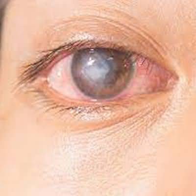
Amebic infection, usually due to Acanthamoeba, is an important cause of keratitis in contact lens wearers.
Although severe pain with perineural and ring infiltrates in the corneal stroma is characteristic, it is not specific and earlier forms with changes confined to the corneal epithelium are identifiable. Diagnosis is facilitated by confocal microscopy. Culture requires specialized media. Longterm treatment is required. Intensive topical biguanide (polyhexamethylene or chlorhexidine) and diamidine (propamidine or hexamidine) is the standard initial treatment with addition of an azole such as voriconazole if necessary. Delayed diagnosis and prior treatment with topical steroids adversely affect the visual outcome. Corneal grafting may be required after resolution of infection to restore vision. If there is scleral involvement, monotherapy or combination therapy with systemic antiinflammatory and immunosuppressant medication is helpful but the prognosis is poor.