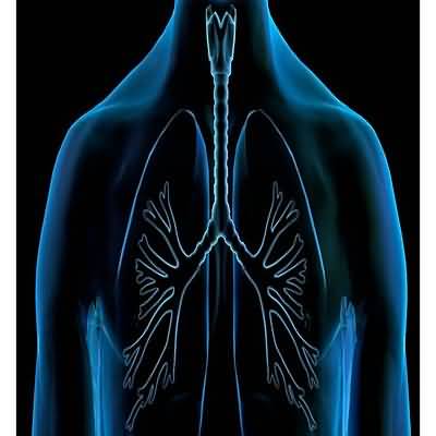
ESSENTIALS OF DIAGNOSISHistory of cigarette smokingChronic cough, dyspnea, and sputum productionRhonchi, decreased intensity of breath sounds, and prolonged expiration on physical examinationAirflow limitation on pulmonary function testing that is not fully reversible and is most often progressive
General Considerations
The Global Initiative for Chronic Obstructive Lung Disease (GOLD) defines COPD as a common, preventable, and treatable disease state characterized by persistent respiratory symptoms and airflow limitation due to airway and alveolar abnormalities usually caused by significant exposure to noxious particles or gases
Symptoms include cough, dyspnea, and sputum production
COPD is a major cause of chronic morbidity and mortality worldwide
Most patients with COPD have features of both emphy- sema and chronic bronchitis
Chronic bronchitis is a clinical diagnosis defined by excessive secretion of bronchial mucus and is manifested by daily productive cough for 3 months or more in at least 2 consecutive years
Emphysema is a pathologic diagnosis that denotes abnormal permanent enlargement of air spaces distal to the terminal bronchiole, with destruction of alveolar walls and without obvious fibrosis
Both of these definitions are no longer included in GOLD because they comprise a minority of patients
Chronic respiratory symptoms also exist in people with normal spirometry, and a number of smokers without airflow limitation will have varying degrees of emphysema
Cigarette smoking is clearly the most important cause of COPD in North America and Western Europe
Nearly all smokers suffer an accelerated decline in lung function that is dose- and duration-dependent
Fifteen percent develop progressively disabling symptoms in their 40s and 50s
Approximately 80% of patients seen for COPD have significant exposure to tobacco smoke
The remaining 20% frequently have a combination of exposures to environ- mental tobacco smoke, occupational dusts and chemicals, and indoor air pollution from biomass fuel used for cooking and heating in poorly ventilated buildings
Outdoor air pollution, airway infection, environmental factors, and allergy have also been implicated in chronic bronchitis, and hereditary factors (deficiency of alpha-1-antiprotease [alpha-1-antitrypsin]) have been implicated in emphysema
Atopy and the tendency for bronchoconstriction to develop in response to nonspecific airway stimuli may be important risks
Evidence suggests that lung exposures to pollution and allergens early in life can lead to poor lung growth in childhood and expiratory airflow limitation, resulting in lower than predicted spirometric values in midlife
Clinical Findings
A
Symptoms and Signs Patients with COPD characteristically present in the fifth or sixth decade of life complaining of excessive cough, sputum production, and shortness of breath
Symptoms have often been present for 10 years or more
Dyspnea is noted initially only on heavy exertion, but as the condition progresses it occurs with mild activity
In severe disease, dyspnea occurs at rest
As the disease progresses, two symptom patterns tend to emerge, historically referred to as “pink puffers” and “blue bloaters”
Most COPD patients have pathologic evidence of both disorders, and their clinical course may involve other factors, such as central control of ventilation and concomitant sleep-disordered breathing
Pneumonia, pulmonary hypertension, cor pulmonale, and chronic respiratory failure characterize the late stage of COPD
A hallmark of COPD is the periodic exacerbation of symptoms beyond normal day-to-day variation, often including increased dyspnea, an increased frequency or severity of cough, and increased sputum volume or change in sputum character
These exacerbations are commonly precipitated by infection (more often viral than bacterial) or environmental factors
Exacerbations of COPD vary widely in severity but typically require a change in regular therapy
B
Laboratory Findings Spirometry provides objective information about pulmonary function and assesses the response to therapy
Pulmonary function tests early in the course of COPD reveal only evidence of abnormal closing volume and reduced midexpiratory flow rates
Reductions in FEV1 and in the ratio of forced expiratory volume to vital capacity (FEV1% or FEV1/ FVC ratio) (Table 9–6) occur later
Post-bronchodilator FEV1/FVC less than 0
70 establishes the presence of airflow obstruction (Global Initiative for Obstructive Lung Disease [GOLD] definition)
In severe disease, the FVC is markedly reduced
Lung volume measurements reveal a marked increase in residual volume (RV), an increase in total lung capacity (TLC), and an elevation of the RV/TLC ratio, indicative of air trapping, particularly in emphysema
Arterial blood gas measurements characteristically show no abnormalities early in COPD other than an increased A–a–Do2
Indeed, measurement is unnecessary unless (1) hypoxemia or hypercapnia is suspected, (2) the FEV1 is less than 40% of predicted, or (3) there are clinical signs of right heart failure
Hypoxemia occurs in advanced disease, particularly when chronic bronchitis predominates
Compensated respiratory acidosis occurs in patients with chronic respiratory failure, particularly in chronic bronchitis, with worsening of acidemia during acute exacerbations
Positive sputum cultures are poorly correlated with acute exacerbations, and research techniques demonstrate evidence of preceding viral infection in a majority of patients with exacerbations
The ECG may show sinus tachycardia and, in advanced disease, chronic pulmonary hypertension may produce electrocardiographic abnormalities typical of cor pulmonale
Supraventricular arrhythmias (multifocal atrial tachycardia, atrial flutter, and atrial fibrillation) and ventricular irritability also occur
C
Imaging Radiographs of patients with chronic bronchitis typically show only nonspecific peribronchial and perivascular markings
Plain radiographs are insensitive for the diagnosis of emphysema; they show hyperinflation with flattening of the diaphragm or peripheral arterial deficiency in about half of cases
CT of the chest, particularly using high-resolution CT, is more sensitive and specific than plain radiographs for its diagnosis
In advanced disease, pulmonary hypertension may be suggested by enlargement of central pulmonary arteries on radiographs, and Doppler echocardiography provides an estimate of pulmonary artery pressure
Differential Diagnosis
Clinical, imaging, and laboratory findings should enable the clinician to distinguish COPD from other obstructive pul- monary disorders, such as asthma, bronchiectasis, cystic Table 9–6
Patterns of disease in advanced COPD
Type A: Pink Puffer (Emphysema Predominant) Type B: Blue Bloater (Bronchitis Predominant) History and physical examination Major complaint is dyspnea, often severe, usually presenting after age 50
Cough is rare, with scant clear, mucoid sputum
Patients are thin, with recent weight loss common
They appear uncomfortable, with evident use of accessory muscles of respiration
Chest is very quiet without adventitious sounds
No peripheral edema
Major complaint is chronic cough, productive of mucopurulent sputum, with frequent exacerbations due to chest infections
Often presents in late 30s and 40s
Dyspnea usually mild, though patients may note limitations to exercise
Patients frequently overweight and cyanotic but seem comfortable at rest
Peripheral edema is common
Chest is noisy, with rhonchi invariably present; wheezes are common
Laboratory studies Hemoglobin usually normal (12–15 g/dL)
Pao2 normal to slightly reduced (65–75 mm Hg) but Sao2 normal at rest
Paco2 normal to slightly reduced (35–40 mm Hg)
Chest radiograph shows hyperinflation with flattened diaphragms
Vascular markings are diminished, particularly at the apices
Hemoglobin usually elevated (15–18 g/dL)
Pao2 reduced (45–60 mm Hg) and Paco2 slightly to markedly elevated (50–60 mm Hg)
Chest radiograph shows increased interstitial markings (“dirty lungs”), especially at bases
Diaphragms are not flattened
